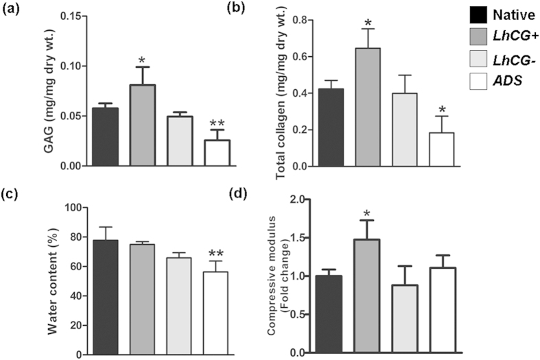
Figure 6. Quantitative evaluation of articular cartilage repair.
Biochemical analysis was performed to quantify the (a) GAG content and (b) total collagen content in all the explanted tissues. To correlate biochemical aspects with the functional aspects of the tissue, (c) water content and (d) compressive moduli were also determined. The results for the three experimental groups, namely LhCG+, LhCG− and ADS group were compared to native, unaffected cartilage. One-way ANOVA/Dunnett’s multiple comparison test was used for data analysis. (NS) indicates no statistical significance between two groups while (*) indicates statistical significance (p < 0.05); (**) (p < 0.01). Data are presented in the form of mean ± SD, with N = 5.

