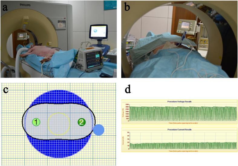
Figure 1. The microsecond-pulsed electric field treatment.
(a) The animal treatment was performed in the interventional radiology suite using a pulse generator and an ECG monitor. (b) The two-needle electrodes were placed with CT scan guidance, and the treatment was performed under general anaesthesia. (c) The two-needle electrodes were placed at a distance predetermined by computer calculation and simulation to bracket the target ablation zone. (d) The electric ablation parameters were 3000 V, 20 A, 90 pulses, and 70 microsecond pulse duration.

