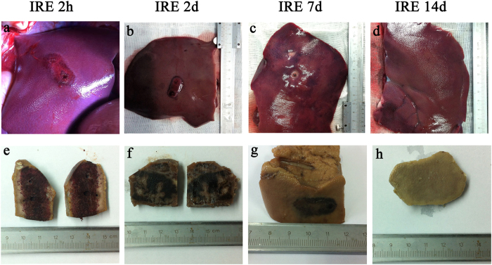
Figure 3. Gross pathologic sectioned specimen of the ablated porcine liver.
Parts (a), (b), (c) and (d) show the discoloration caused by ablation on the dissected liver 2 hours, 2 days, 7 days and 14 days post-treatment, respectively. Parts (e), (f), (g) and (h) show the sharp demarcation of the ablated zone on the formalin-fixed liver. Intact vessels and bile ducts are seen in the area of ablation. The ablated zone showed vascular congestion and haemorrhagic change, with grossly intact hepatic morphology, 2 hours, 48 hours and 7 days after treatment. By day 14, the areas of vascular congestion and haemorrhage had resolved in the ablated zone. A well-demarcated margin was visualized between the ablated and non-ablated zones. The traversing vessels and bile ducts appeared intact within the ablated zones.

