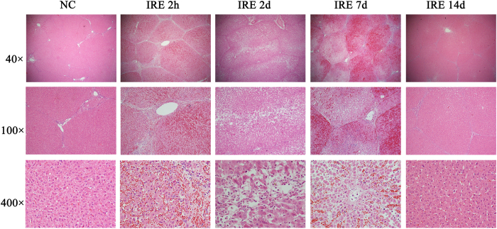
Figure 4. Haematoxylin and eosin (H&E) stained sections.
Figure 4 shows haemorrhagic change with relatively intact hepatic morphology. Each hepatic lobule was visible and intact throughout the ablation area. The vessels and bile ducts appeared to have mild oedema without apparent structural destruction. On the 2-hour, 2-day and 7-day H&E slides, there was acute, extensive, and severe cell death. No viable cells were detected within the ablated area. However, the normal hepatic architecture was preserved. The ablation creates three layers: in the centre layer, where the electrode was inserted, there is haemorrhaging and severe necrosis; in the middle layer, there is coagulative type cell death; and in the outer layer, there are clusters of cell death. From 2 days to 7 days, the ablation area was filled with congestion from neutrophil and eosinophil infiltration. The larger vessels and bile ducts in the ablated area appeared structurally preserved. However, there was mild vaculitis, multifocal loss of endothelial integrity, oedema, separation of the tunica muscularis layers, and neutrophilic infiltration. The bile ducts showed signs of acute choledochitis with peridochal oedema. After 14 days, there was extensive hepatocellular regeneration in the ablation area.

