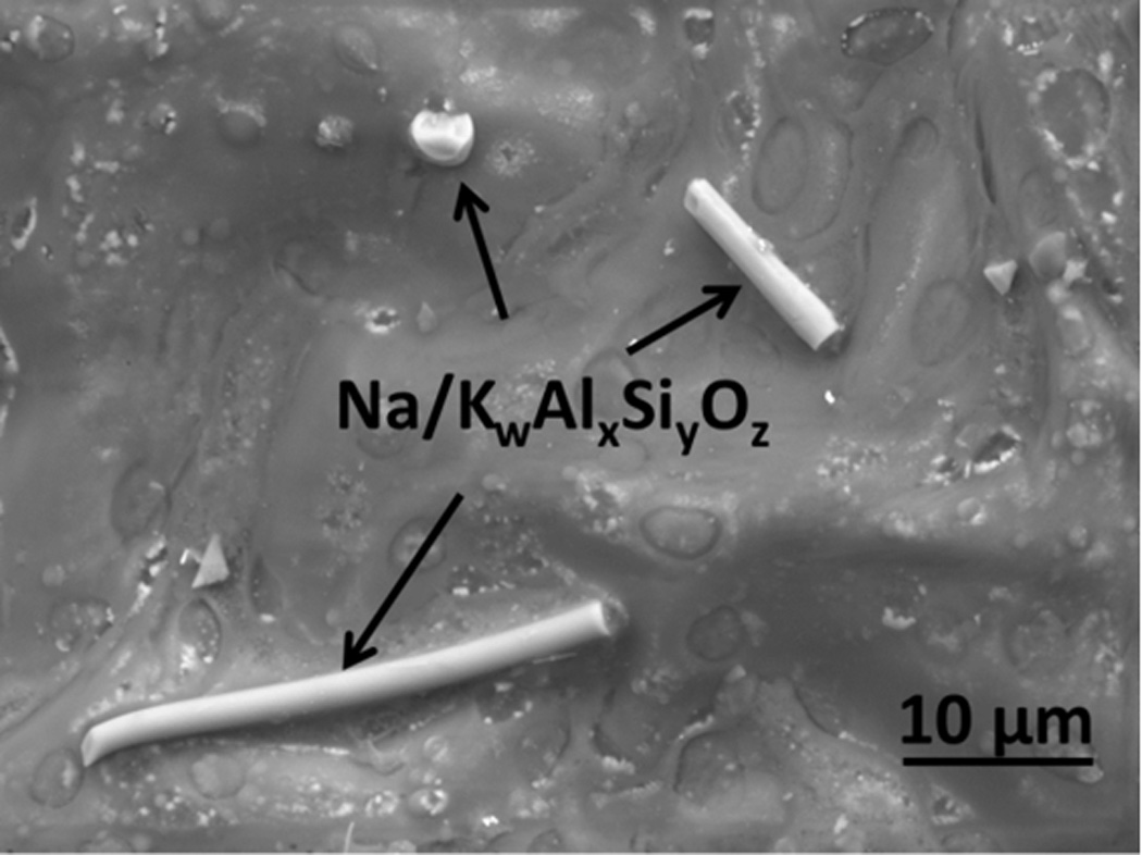
Figure 1.

A SEM micrograph of soil halloysite aluminum silicate crystals on a popular commercial chewing tobacco leaf surface. The particle at the top of the image is also an aluminum silicate, though not identifiable on the basis of morphology. This image was obtained in Environmental (ESEM) mode with an accelerating voltage of 10kV, spot size 5.0, 2 torr vapor pressure in the sample chamber, with a 500µm pressure limiting aperture on a Gaseous Analytical Detector (GAD).
