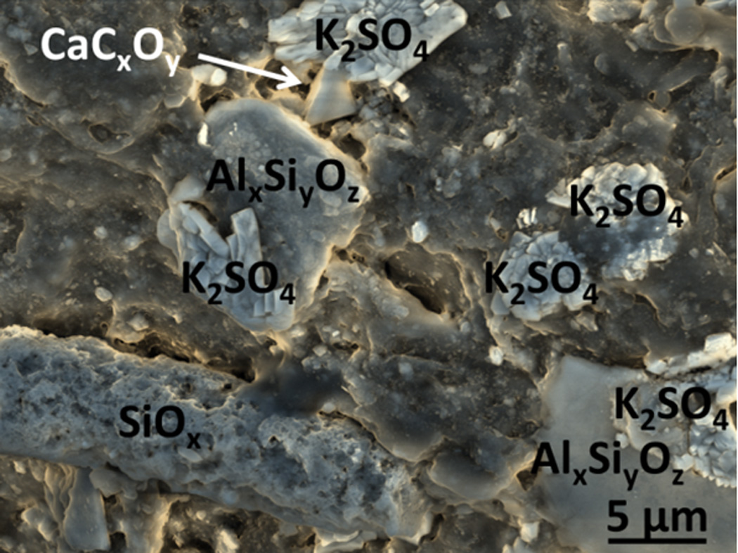
Figure 3.

False color SEM micrograph of particles on the surface of reconstituted cigarette filler tobacco from a popular cigarette brand. Two aluminum silicate particles of different sizes are visible, along with potassium sulfate crystals, and apparently calcium oxalate. The elongated structure is a halloysite-like silica structure with numerous etch pits, but negligible aluminum. This image was obtained in Low Vacuum mode with an accelerating voltage of 10 kV, 0.6 torr vapor pressure in the sample chamber, spot size 3.0, and mixed Large Field (LFD) and Concentric Backscatter Detectors (CBS).
