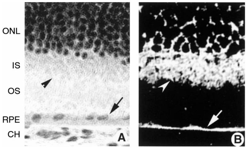
Fig. 2.
Laser scanning confocal microscopic immunolocalization of taurine transporter in intact mouse retinal tissue. Eyes of ICR albino mice were frozen in OCT embedding medium, and cryosections were prepared and subjected to immunohistochemical detection of taurine transporter using a commercially available antibody. A: hematoxylin-and-eosin-stained cryosection of outer portion of normal mouse retina showing outer layers of the retina. B: immunolocalization of taurine transporter. Arrow points to intense labeling of the RPE; arrow-head points to labeling of inner segments (IS). Magnification ×400. ONL, outer nuclear layer; OS, outer segments; CH, choroid.

