Figure 4.

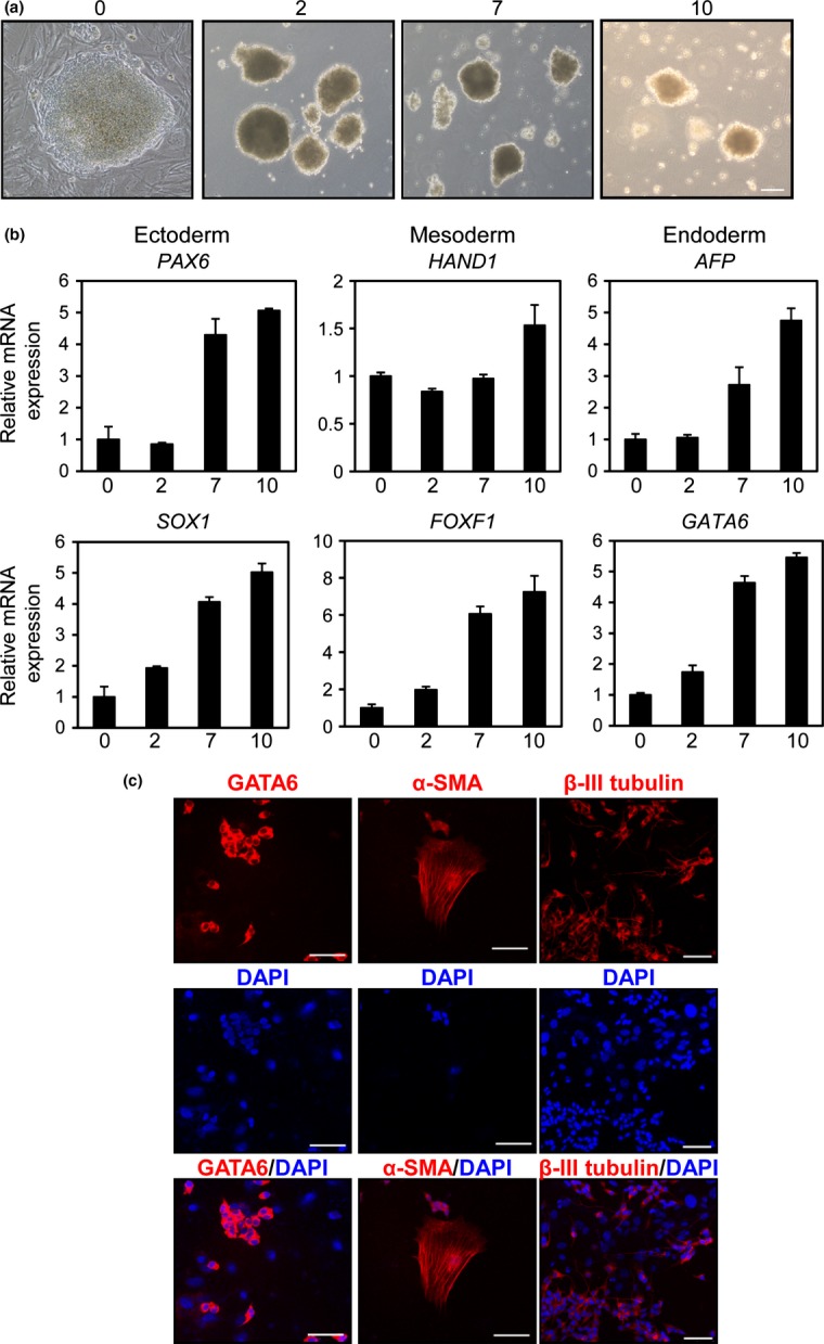
Embryoid body (EB)-mediated differentiation of SH-IN 4F cells. (a) Representative phase contrast images of EBs generated from SH-IN 4F cells. Undifferentiated cells at day 0 and differentiated EBs at days 2, 7, and 10. Scale bar: 100 μm. (b) Expression of ectodermal (PAX6 and SOX1), mesodermal (HAND1 and FOXF1) and endodermal (AFP and GATA6) markers were examined by qPCR. The x-axis represents each time point in days and the y-axis represents relative fold induction compared with day 0. All values were normalized to β-actin mRNA expression levels. (c) Immunofluorescence analysis of SH-IN 4F (clone 2) after EB differentiation: endoderm (GATA6), mesoderm (α-smooth muscle actin; [α-SMA]) and ectoderm (βIII tubulin). Nuclei were stained with DAPI (blue). Scale bars: 75 μm (GATA6 and α-SMA) and 50 μm (βIII tubulin). Results are representative of three independent experiments.
