Figure 5.

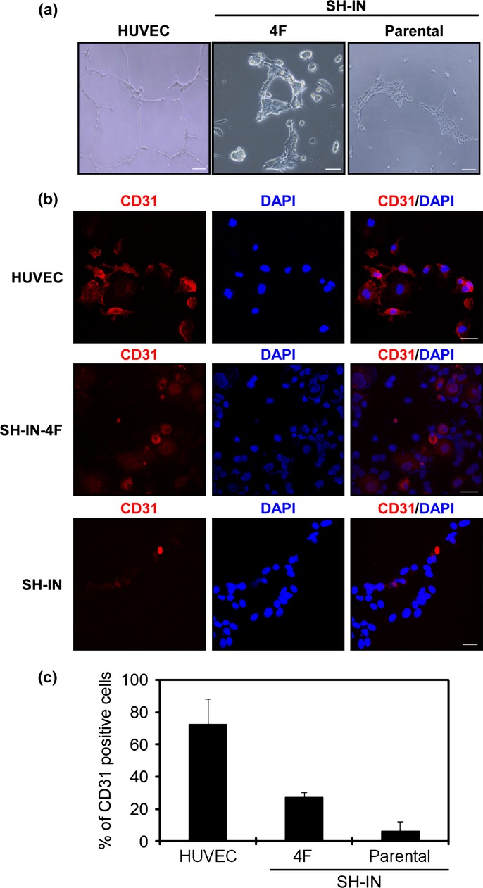
Endothelial tube formation by SH-IN 4F cells. (a) SH-IN 4F cells cultured in serum-free medium supplemented with basic fibroblast growth factor (bFGF) and EGF to examine plasticity. Representative micrograph of the tube network formed by SH-IN 4F cells and HUVEC cells. Scale bar: 300 μm. (B) HUVEC cells or SH-IN cells were cultured in differentiating medium supplemented with 2% FCS and vascular endothelial growth factor (VEGF for 7 or 10 days, respectively. Expression of the endothelial-specific marker CD31 was detected by immunofluorescence. Scale bar: 50 μm. Nuclei are stained with DAPI (blue). (c) Quantification of CD31-expressing cells as measured by immunofluorescence. Columns represent mean values from three replicate experiments, error bars represent SD. Results are representatives of three independent experiments performed using clone 7.
