Figure 6.

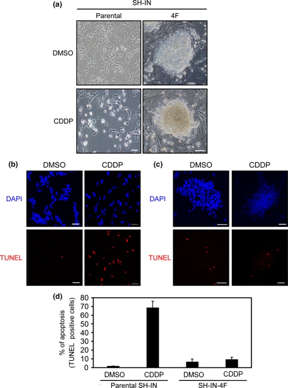
Reprogramming of SH-IN cells resulted in chemoresistance. (a) Brightfield micrographs of SH-IN cells (clone 2) treated with DMSO (top left) or 20 μM CDDP (bottom left) for 12 h. All images were acquired with an Olympus microscope using a 10 × objective lens. Scale bar: 100 μm. SH-IN (b) and reprogrammed (c) SH-IN 4F cells were treated with DMSO and CDDP (20 μM) for 12 h and subjected to TUNEL assays (red). Cell nuclei were stained with DAPI (blue). Scale bar: 50 μm. (d) TUNEL-positive cells from three independent experiments. Values are presented as the mean ± SD. Results are representatives of three independent experiments.
