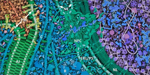
Fig. 1.
An example to illustrate the crowded nature of the cell. Here, two adjacent vascular endothelial cells are shown connected via an adherens junction (AJ) between the opposing plasma membranes (PM) of the two cells. The adherens junction contains localized clusters of cell adhesion proteins, mainly cadherins and catenins (green). Note that other plasma membrane proteins such as vascular endothelial growth factor receptors (yellow) are excluded from the clusters. Extracellular blood plasma proteins are shown in tan (upper left). Cytoplasmic proteins are shown in turquoise, and nuclear proteins are shown in purple and blue (right). The image also displays common features of the eukaryotic cell, including ribosomes (R), cytoskeletal cables (C), the nuclear pore complex (NPC), the nuclear lamina (NL), the nuclear envelope (NE), and the ER lumen/perinuclear space (ERL/PS). The macromolecules are shown at their approximate true densities and molecular dimensions. For more details about the biological context of this image, see Span et al. (73) (used with permission of David S. Goodsell, the Scripps Research Institute).

