Fig. 5.
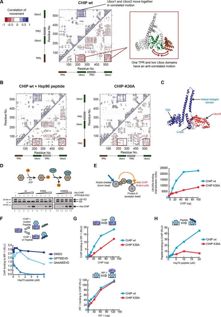
Coordinated movements between the TPR and U-box regulate CHIP activity. (A) Dynamic cross-correlation map (left panel) of Cα atoms for the un-liganded wt CHIP dimer. Correlated motions are represented above the diagonal in blue and anticorrelated below in red. Correlated movements of the CHIP U-boxes are indicated by a blue box. Anticorrelated movements of the TPR domain (right panel in brown) with both U-boxes (right panel in green) are indicated with red boxes. Cartoon of CHIP dimer (right panel) was generated using PyMOL v.1.4.1. (B) As above except the dynamic cross-correlation maps of Cα atoms are for Hsp90 peptide bound wt CHIP dimer (left panel) and the K30A mutant CHIP dimer (right panel). (C) Snapshot of the crystal structure of zebrafish CHIP-Ubox in complex with UbcH5 (from PDB 2OXQ) superimposed onto the crystal structure of mouse CHIP (from PDB 2C2L). The image, showing a single CHIP monomer, was generated using PyMOL v1.4.1. Blue ribbon: CHIP; red ribbon: UbcH5. (D) His-UbcH5a was charged with ubiquitin (Ub∼E2; thiolester linkage) by incubating with UBE1 and ubiquitin in the presence of ATP, following which ubiquitin discharge from the E2 by His-CHIP wt or K30A mutant was monitored. The E2-binding-defective mutant H260Q was included as a control. Shown is an immunoblot probed for CHIP and the E2. (E) An AlphaScreen assay was set up (see cartoon) to measure binding dynamics of untagged CHIP wt or K30A mutant anchored on protein A acceptor beads with His-tagged UbcH5a captured on Nickel-chelate donor beads in solution. (F-G) GST alone controls showed negligible binding and are therefore not indicated on the graphs. (F) Binding assay with fixed amounts of GST-IRF-1 immobilized on microtitre wells. Fixed amounts of His-CHIP wt together with a carrier control (DMSO) or a titration of Hsp70 wt or mutant peptide was added in the mobile phase. CHIP binding to IRF-1 was measured on a luminometer using an anti-CHIP antibody. (G) Upper panel: Binding assay as in (F) except that a titration of His-CHIP wt or K30A mutant was added in the mobile phase. Lower panel: Binding assay with fixed amounts of His-CHIP wt or K30A mutant coated on microtitre wells and a titration of GST-IRF-1 added in the mobile phase. (H) Binding assay as in (G) (lower panel) except that a titration of Hsp70 wt peptide was added in the mobile phase. Binding was detected using streptavidin-HRP.

