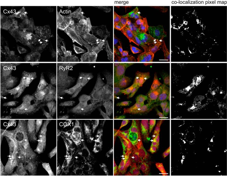
Fig. 6.
RyR2 and COX1 colocalize with Cx43 in HL-1 cells. Immunostaining for Cx43 (Sc9059) and RyR2 (middle panel) and COX1 (lower panel), in HL-1 cells. Positive control was performed by colocalization analysis with F-actin, stained with Phalloidin (upper pannel, Cx43 staining with 610062). Nuclei were stained with DAPI. Scale bars, 20 μm. Arrows indicate colocalization spots. Colocalization pixel map is depicted on the right side.

