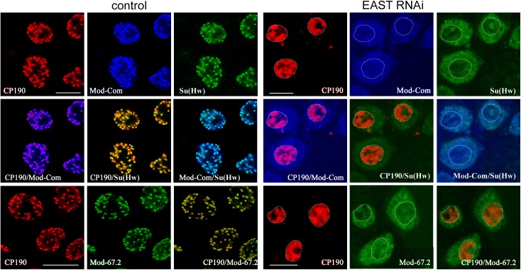
Fig 3. Immunofluorescent localization of insulator proteins in the nuclei of S2 cells after EAST knockdown.
EAST RNAi was performed with two sets of primers: one specific for the 5’ end of east transcript (5’ cgtcggcgaagagatgtcta 3’ and 5’ ttgctctgttactgaggaggatgca 3’) and the other, for the middle part of east transcript (5’ taagtcaagcgggaccttgg and 5’ atcccgctgcagaccccat 3’). Nontransfected S2 cells designated as «control» and S2 cells after EAST knockdown by RNAi designated as «EAST RNAi». Immunostaining with antibodies to CP190 (red), Su(Hw) (green), Mod(mdg4)-67.2 (Mod-67.2, green), and common part of Mod(mdg4) (Mod-Com, blue). Dotted line indicates the nucleus boundary (See S5 Fig). Scale bars, 5 μm.

