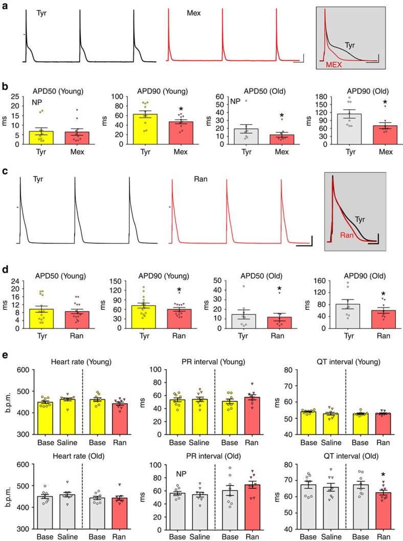
Figure 4. Inhibition of INaL shortens the repolarization properties of the old heart.
(a) APs recorded in an old myocyte before (Tyrode, black traces) and after exposure to the Na+ channel inhibitor mexiletine (Mex, red traces). Scale bars, 200 ms, 20 mV. Superimposed traces are reported in the inset. Scale bars, 500 ms, 20 mV. (b) Quantitative data in myocytes from male mice at 3 months (Young, n=10 cells from 5 hearts) and 27–30 months (Old, n=8 cells from 5 hearts) before (Tyr) and after exposure to 10 μM mexiletine (Mex) are shown as mean±s.e.m. and scatter plots. Tyr, Tyrode solution. *P<0.05 versus Tyr (paired t-test and Wilcoxon signed rank test); NP, non-parametric analysis. Additional parameters are reported in Supplementary Fig. 18. (c) APs recorded in an old myocyte before (Tyrode, black traces) and after exposure to INaL inhibitor ranolazine (Ran, red traces). Scale bars, 300 ms, 20 mV. Superimposed traces are reported in the inset. Scale bars, 50 ms, 20 mV. (d) Quantitative data in myocytes from mice at 3–6 months (Young, n=14 cells from 4 hearts) and 27–33 months (Old, n=8 cells from 5 hearts) before (Tyr) and after exposure to 10 μM ranolazine (Ran) are shown as mean±s.e.m. and scatter plots. *P<0.05 versus Tyr (paired t-test). Additional parameters are reported in Supplementary Fig. 18. (e) Quantitative data for electrocardiographic parameters obtained in male mice at 3–4 months (Young) and at 30–31 months (Old) at baseline (Base) and 1 h after treatment with saline (Young, n=9; Old n=8) or ranolazine (30 mg per kg body weight, i.p.) (Young, n=8; Old n=8). Data are shown as mean±s.e.m. and scatter plots. *P<0.05 versus Base (paired t-test and Wilcoxon signed rank test); NP, non-parametric analysis.

