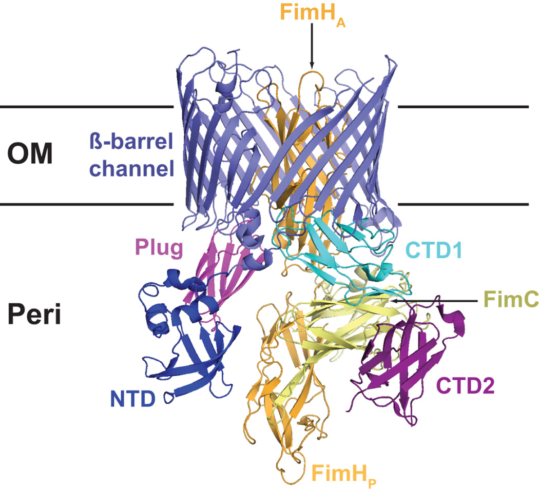
Figure 3.
Crystal structure of the FimD-FimC-FimH type 1 pilus assembly intermediate (PDB ID: 3RFZ). The Usher NTD, plug, β-barrel channel, and CTD domains are indicated. The FimH adhesin domain (FimHA) is inserted inside the usher channel, and the FimH pilin domain (FimHP) and bound FimC chaperone are located at the usher CTDs.

