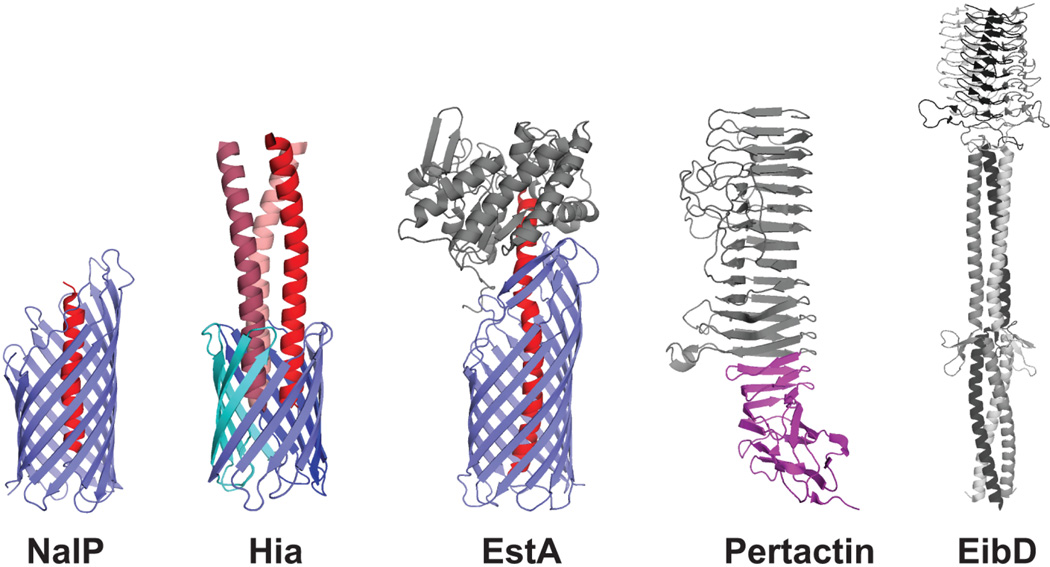
Figure 6.
Crystal structures of representative autotransporter proteins. Translocator domains from the monomeric NalP and trimeric Hia autotransporters are shown (PDB IDs: 1UYN and 2GR7, respectively), with the β-barrel channels in blue and the α-helical linker regions in red. Passenger domains from the monomeric Pertactin and trimeric EibD autotransporters are shown (PDB IDs: 1DAB and 2XQH, respectively), with the approximate location of the Pertactin autochaperone region indicated in purple. The complete structure of the EstA autotransporter is shown (PDB ID: 3KVN), with the translocator domain in blue, the α-helical linker in red, and the globular passenger domain in gray.

