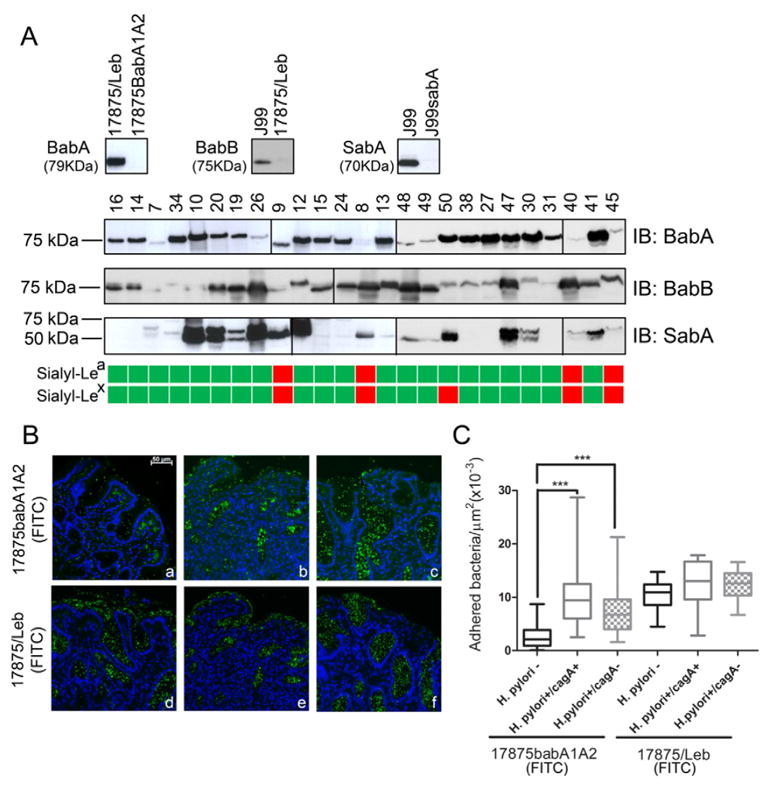
Fig. 5.

Impact of increased sialylation on H. pylori attachment to inflamed human gastric mucosa. (A) Immunoblot analysis of model H. pylori strains and clinical isolates using BabA, BabB and SabA recognizing antibodies, and schematic representation of sialyl-Lea and sialyl-Lex expression in the corresponding individuals gastric biopsies (green: positive; red: negative). (B) Adhesion of fluorescein-labeled H. pylori strain 17875babA1A2 (functional SabA+) and 17875/Leb (functional SabA −) to gastric mucosa tissue sections from non-infected (H. pylori−) (a, d), H. pylori CagA(+) strains infected (H. pylori+/CagA+) (b, e) and H. pylori CagA(−) strains infected (H. pylori+/CagA−) (c, f) individuals, magnification 200×. (C) Quantification of bacterial adhesion to human gastric mucosa tissue samples, the box and whisker plots represent the minimum and maximum values and the median of at least 24 different fields from three independent gastric biopsies for each biological group; significance was determined by one-way ANOVA with Dunnett’s Multiple Comparison Test. ***p < 0.001.
