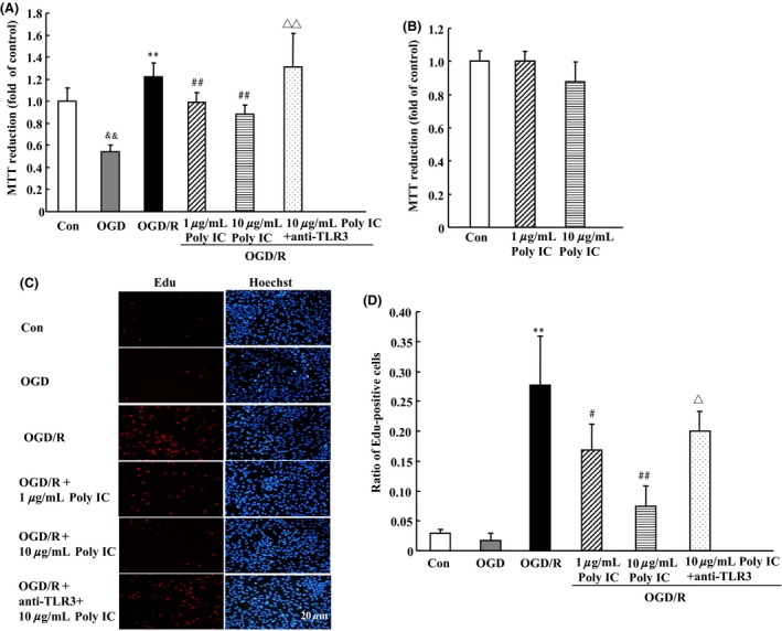
Figure 1.

Poly IC inhibits oxygen–glucose deprivation/reoxygenation (OGD/R)‐induced astrocyte proliferation. Cells were exposed to OGD for 6 h followed by reoxygenation for 24 h. Poly IC (1 and 10 μg/mL) or saline was applied to the media at the beginning of reoxygenation. (A and B) Cell viability was measured by the MTT assay. The control group (Con) was grown in standard medium under normoxic conditions. (C) Cell proliferation was assayed by EdU immunostaining. Scale bar = 20 μm. (D) Statistical analysis of the percentage of EdU‐positive astrocytes in each group. Results are expressed as mean ± SD and are from three independent experiments. && P < 0.01 versus control group; **P < 0.01 versus OGD group; # P < 0.05, ## P < 0.01 versus OGD/R group; Δ P < 0.05, ΔΔ P < 0.01 versus 10 μg/mL Poly IC‐treated group.
