Fig. 4.

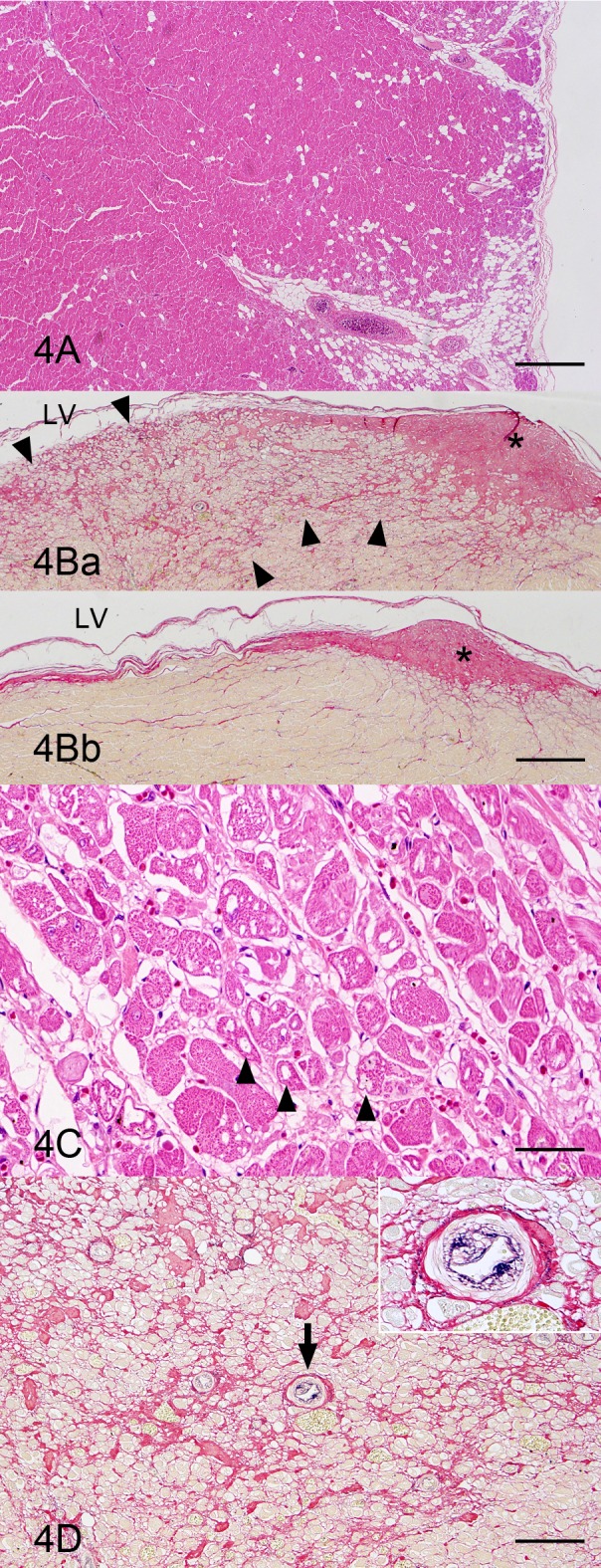
Heart; left ventricular wall; a boxer dog with ARVC. (A) Loss and fibrofatty replacement of cardiocytes are observed in the outer muscle layer. As compared to the right ventricular wall (Fig. 3A), the lesion is limited. HE stain. Bar=1 mm. (B) Fibrosis is remarkable in the papillary muscle region (Fig. 4Ba) compared to the intact region of this animal (Fig. 4Bb). The fibrosis (arrowheads) occurs apart from a tendinous cord (asterisk). LV, left ventricle. Modified elastica van Gieson stain. Bar=500 µm. (C) The cardiocytes having vacuoles are distributed in the papillary muscle region where fibrosis is observed. HE stain. Bar=50 µm. (D) The intimal thickening of small arteries (arrow and inset) at the fibrosis region in the papillary muscle is evident. Modified elastica van Gieson stain. Bar=100 µm.
