Fig. 5.

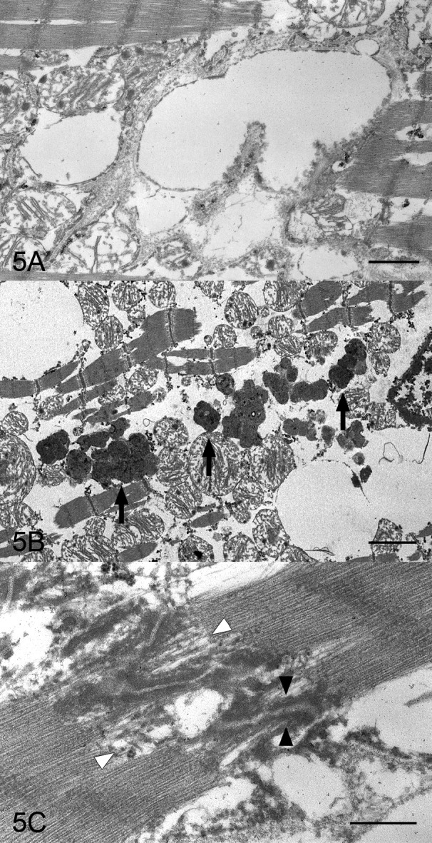
Heart; right ventricular wall; a boxer dog with ARVC. (A) The cardiocyte of outer muscle layer has some vacuoles which are surrounded by a single layer membrane. Electron micrograph. Bar=1 µm. (B) Many high electron dense materials (arrows) corresponding to lipofuscins on light microscopy are in the cardiocytes. Electron micrograph. Bar=2 µm. (C) The intercalated disk in the cardiocytes is of a high electron density (black arrowheads), and the myofibrils come loose around the intercalated disks (white arrowheads). Electron micrograph. Bar=0.5 µm.
