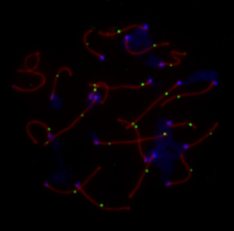
Fig 1. Representative image of a late pachytene spermatocyte stained with fluorescently labeled antibodies.
SCP3, a component of the lateral elements of the synaptonemal complex, is stained in red. Sites of crossing over along the synaptonemal complex are denoted by green MLH1 foci. Centromeric proteins targeted by CREST antibodies are in blue. The white arrow points to the heterogametic sex chromosomes. Only MLH1 foci on autosomal bivalents were scored in this study (n = 23 for this image).

