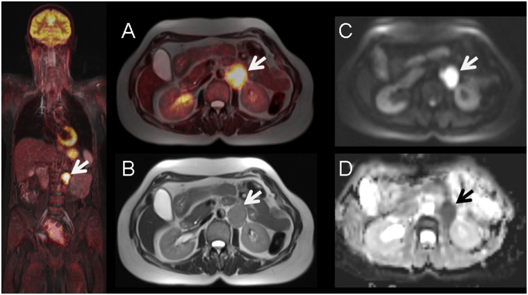
Fig 1. 56-year old female with histology proven recurrent lymph node metastasis of cervical cancer diagnosed 3 years before.
Pre-operative simultaneous 18F-FDG-PET/MRI (A) and T2-weighted MR imaging (B) show a hypermetabolic left paraaortal metastastic lymph node (arrow) with corresponding diffusion-restriction in DWI (C) and ADC-map (D).

