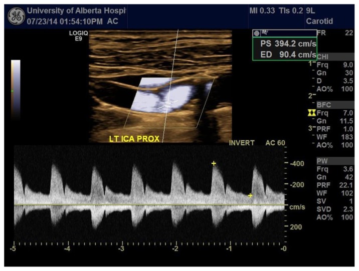
Figure 4.
A 76-year-old male with an incomplete web in the left proximal internal carotid artery. FINDINGS: Spectral Doppler ultrasound of the neck demonstrates stenosis of the left proximal ICA due to the carotid web. There is a markedly elevated peak systolic velocity on spectral Doppler. TECHNIQUE: Spectral Doppler imaging was performed on a GE Logiq E (xd-clear platform) equipped with a linear array transducer (9L probe) at the carotid preset, frequency = 3.6 MHz, wall filter = 96. The pulsed wave Doppler was positioned over the vessel to be interrogated. The gain was set to 42 for optimal vessel delineation free of noise. Beam steering and angle correction were performed to acquire accurate flow measurements, and the pulse repetition frequency was set to 20.9 Hz for optimal Doppler sampling.

