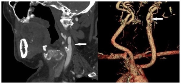
Figure 5.
A 76-year-old male with an incomplete web in the left proximal internal carotid artery. FINDINGS: Sagittal contrast-enhanced CT of the neck during the arterial phase and three dimensional volume rendered CT angiography of the carotid arteries show an incomplete web (arrow) in the left proximal ICA, causing >70% stenosis. TECHNIQUE: Sagittal CT of the neck, tube current = 486 mAs, tube voltage = 120 kV, slice thickness = 3 mm, intravenous administration of 75 mL Omnipaque 350 contrast material at a flow rate of 5–6 mL/s.

