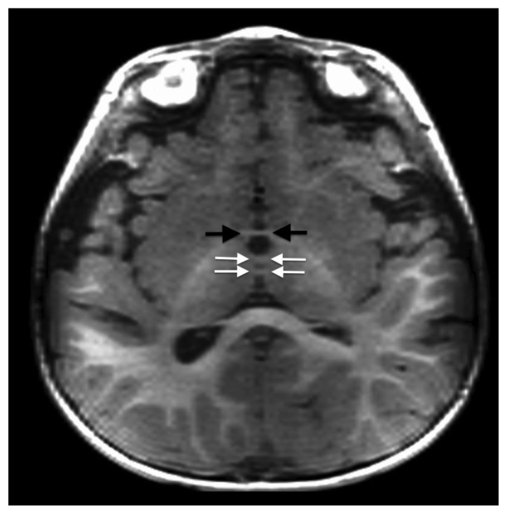
Figure 2.
14-month-old female with duplication of the massa intermedia. FINDINGS: axial T1WI demonstrates 2 distinct horizontally oriented parenchymal bands crossing the 3rd ventricle connecting the thalami consistent with massa intermedia duplication (white arrows). The normal anterior commissure is visible anteriorly (black arrows). TECHNIQUE: 1.5T MR (General Electric, Milwaukee, WI). Fast Spoiled Gradient Echo (FSPGR) image (TR/TE/IT = 11/2/500).

