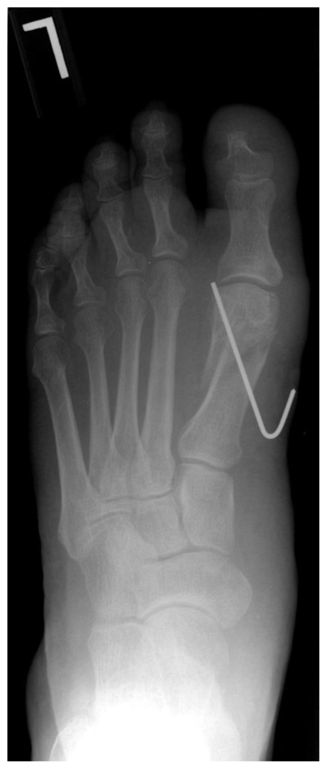
Figure 10.
This AP post-operative radiograph demonstrates a distal first metatarsal procedure fixated with a percutaneous K-wire. This is an acceptable form of fixation and is typically removed after a period of several weeks. Proper position of the K-wire entails that it is free of adjacent joints and avoids soft tissue structures. (Image courtesy of Jane Pontious, DPM FACFAS)

