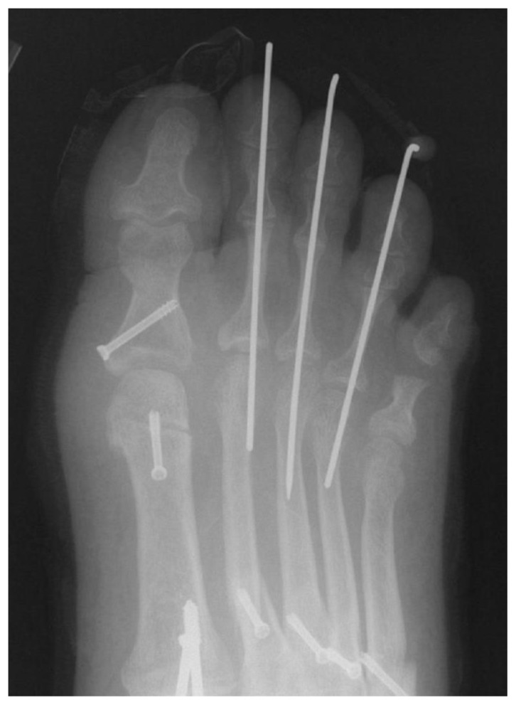
Figure 13.
Post-operative weight-bearing AP radiograph of the 28 y/o patient in Figure 12 following a Reverdin osteotomy within the head of the first metatarsal (among other procedures). Note the realignment of the articular cartilage on the head of the first metatarsal in line with the long axis of the first metatarsal shaft compared to the pre-operative view.

