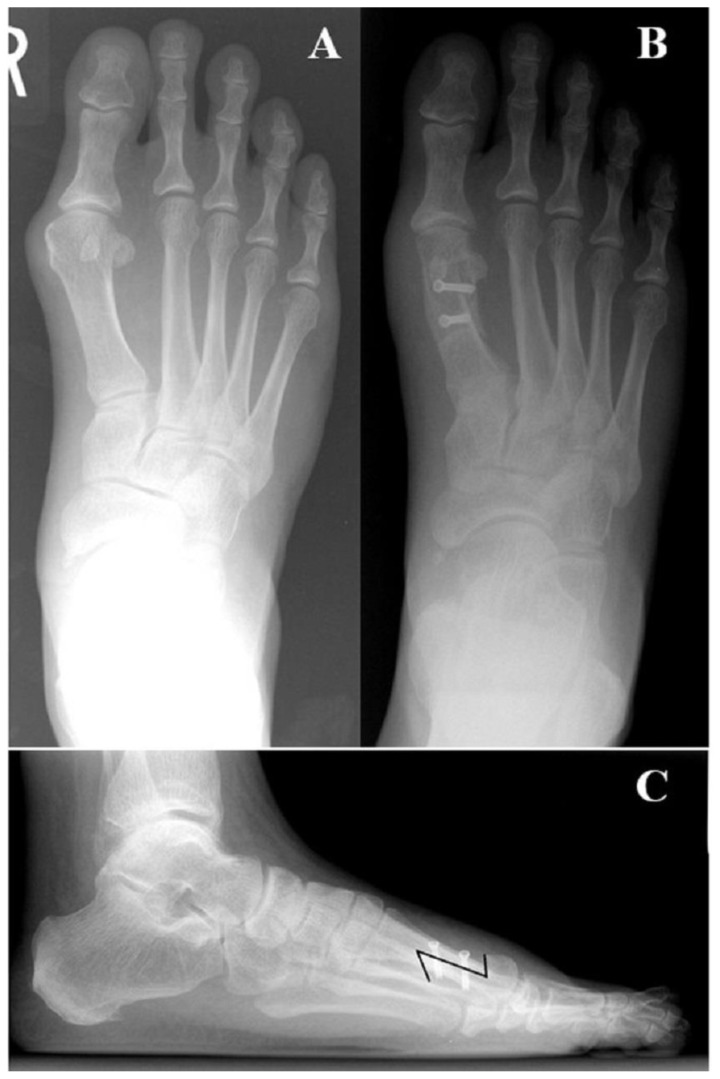
Figure 17.
This series of images illustrates the surgical correction of a HAV deformity in a 63 y/o male patient. Figure 17a represents the pre-operative weight-bearing AP view of the right foot with apparent increases in the 1st IMA (approximately 15 degrees), HAA (approximately 23 degrees) and MSP (approximately 5). These findings are consistent with a moderate HAV deformity. Figure 17b is the post-operative weight-bearing AP view which demonstrate reduction of radiographic parameters to their normal ranges. Figure 17c shows the post-operative weight-bearing lateral view where appropriate orientation and length of the bicortical screws can be appreciated.

