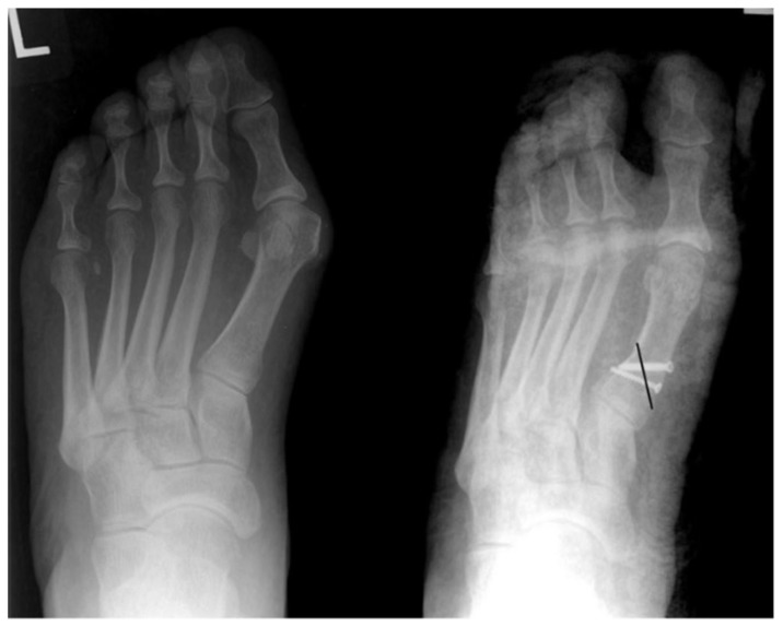
Figure 19.
These pre- and post-operative weight-bearing AP radiographs demonstrate the surgical correction of a HAV deformity with a closing base wedge osteotomy in a 49 y/o female patient. A black line has been superimposed on the post-operative image to demonstrate the location and orientation of the osteotomy. One can appreciate that two screws have been positioned across the osteotomy where the laterally based wedge was resected and closed down. The lateral aspect of the shaft of the first metatarsal now shows a small “step-off” which is consistent with a closing base wedge osteotomy. (Radiographs courtesy of Dr. Joshua Moore, DPM.)

