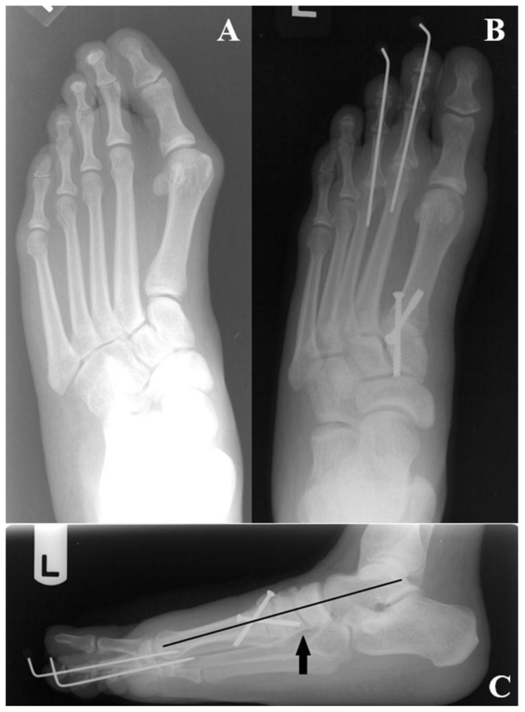
Figure 21.
Pre- and post-operative radiographs of a 23 y/o female with a HAV deformity corrected with a first metatarsal-medial cuneiform arthrodesis (among other hammertoe procedures.) Figure 21a represents the pre-operative weight-bearing AP view of the left foot with increases in the 1st IMA, HAA and MSP which have been reduced to within their normal ranges on the post-operative weight-bearing AP view in Figure 21b. In fact, one can appreciate the near parallel relationship between the first and second metatarsals on the post-operative view. The arthrodesis has been fixated with two crossing screws at metatarsal-cuneiform joint level, with two K-wires used for fixation of the hammertoes of digits two and three. On the post-operative lateral view (Figure 21c), note the parallel relationship between the longitudinal axes of the talus and first metatarsal. Also note that the distal-to-proximally oriented screw appears to penetrate the navicular-cuneiform joint on the AP view, but is appreciated to be clear of this joint when viewed from the lateral projection (see arrow).

