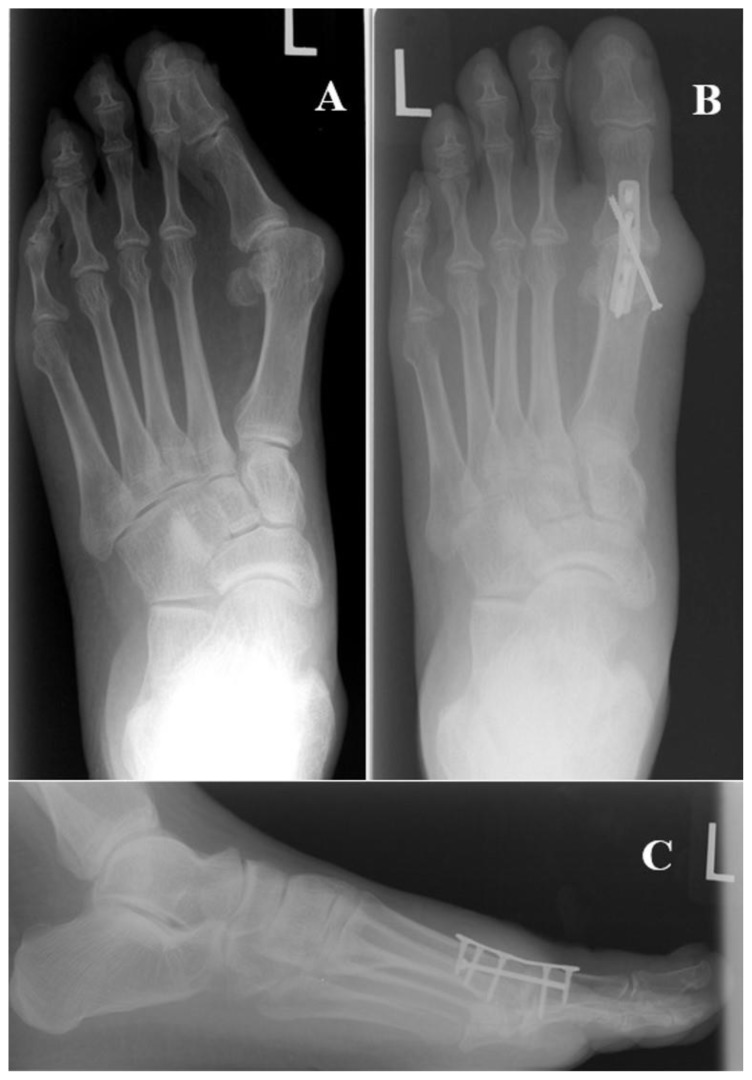
Figure 24.
These figures demonstrate pre- and post-operative weight-bearing AP and lateral radiographs of a 66 y/o male patient who underwent first metatarsal-phalangeal arthrodesis. Pre-operatively in Figure 24a we note a significant deformity with increases in the 1st IMA (approximately 19 degrees here), HAA (approximately 39 degrees here) and MSP (approximately 7 here), but significant return to normal range post-operatively (Figure 24b). This procedure is usually reserved for patients with significant degenerative joint disease or an unstable joint with rheumatoid arthritis, as was the case with this patient. The plate utilized for this procedure is pre-contoured to assist with post-operative joint position. Surgeons attempt to place the joint in approximately 10 degrees of dorsiflexion, 10 degrees of abduction and 0–5 degrees of valgus.

