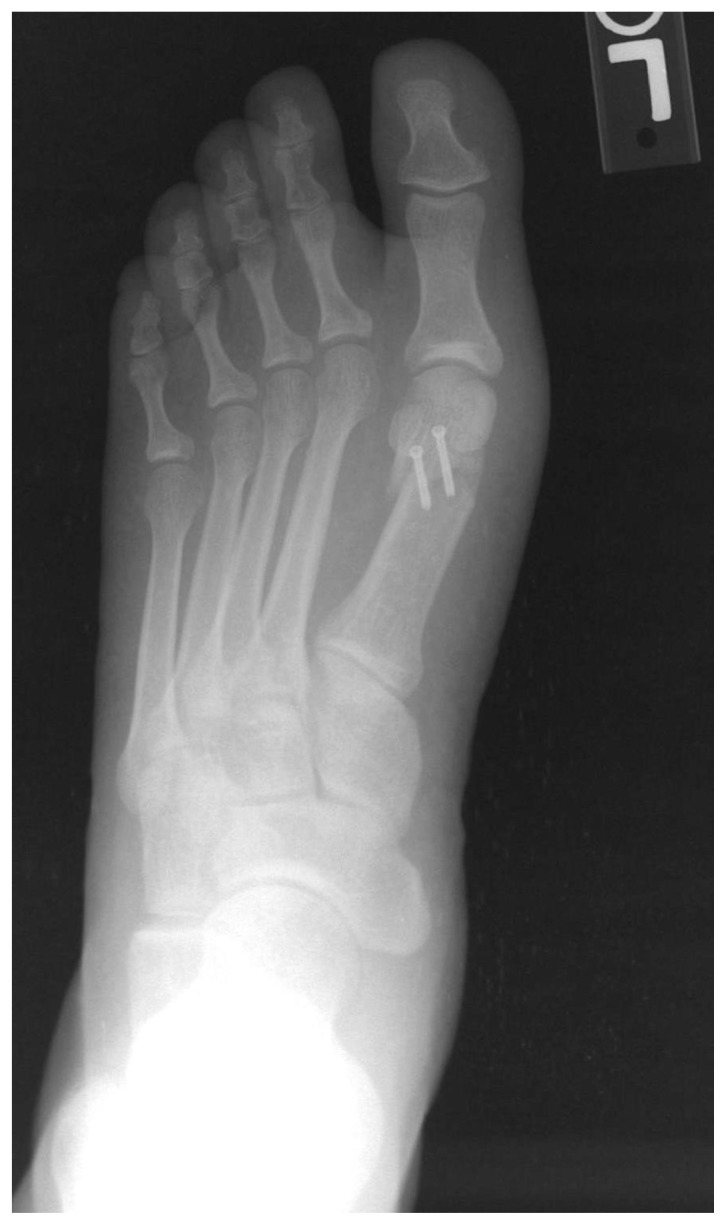
Figure 6.
Post-operative AP radiograph of the 34 y/o female from Figure 1 status post surgical correction of the deformity. One can appreciate reduction of the 1st IMA, HAA and MSP to within normal ranges. Note how the medial aspect of the tibial sesamoid is now in complete alignment with the medial surface of the first metatarsal. Additionally, two parallel screws can be appreciated crossing the osteotomy within the first metatarsal head. These radiographic findings are consistent with appropriate post-surgical changes following distal first metatarsal osteotomy for correction of the HAV deformity.

