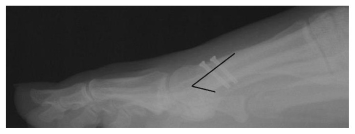
Figure 7.
Post-operative weight-bearing lateral radiograph of the 34 y/o female from Figure 1 status post surgical correction of the deformity. On this lateral view, one can appreciate that two bicortical screws were inserted perpendicular to the osteotomy and angled away from the metatarsal-sesamoid articulation. One can also appreciate appropriate length on these two screws with several thread lengths penetrating the plantar cortex of the metatarsal. The black line on the image emphasizes the location and shape of the performed osteotomy.

