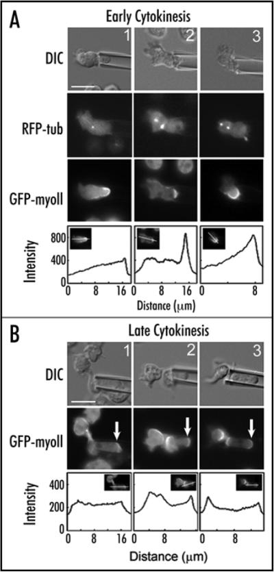
Figure 2.
Myosin-II redistributes to the site of cell deformation during anaphase through the end of cytokinesis. (A) Three examples of mitotic cells aspirated during anaphase. DIC images show the cell and the micropipette. RFP-tubulin images reveal anaphase spindles that are often bowed during the cell deformation. GFP-myosin-II becomes enriched under the micropipettes. Line scans show the increased GFP-myosin-II accumulation at the micropipette. (B) Three examples of late stage dividing cells aspirated by the micropipette (DIC). GFP-myosin-II has accumulated at the micropipette (arrows) and at the cleavage furrow cortex. Line scans show the increased GFP-myosin-II accumulation at the micropipette. Scale bars in (A and B) are 10 mm and apply to all panels.

