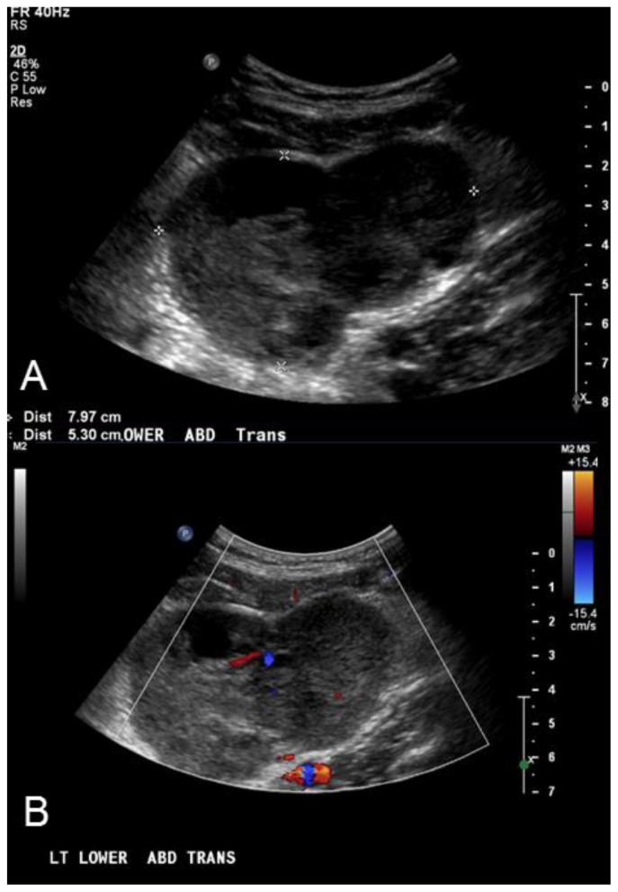
Figure 1.
27 year old male with Desmoplastic Small Round Cell Tumor (DSRCT).
Findings: A) Ultrasound imaging of the abdomen demonstrates a midline heterogeneous, hypoechoic, lobular 8 × 5.3 × 6.6 cm mass (outlined by white cursors). B) Color Doppler ultrasound of the mass in the left lower abdomen demonstrates the present of flow within the hypoechoic mass, indicating a solid lesion.
Technique: Abdominal Ultrasound with Curvilinear 5-2 MHz probe.

