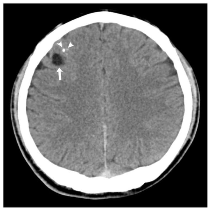
Figure 1.
25-year-old male with angiocentric glioma in the right frontal lobe.
Findings: Noncontrast axial CT scan demonstrates a low density lesion (arrow) involving the right frontal region. There is a small area of calcification (CT value=116 HU) located anterior to the lesion (arrowhead).
Technique: Siemens (SOMATOM Emotion) 16-slice multidetector CT, sequential axial, 270 mAs, 130 kVp, 4.8 mm slice thickness.

