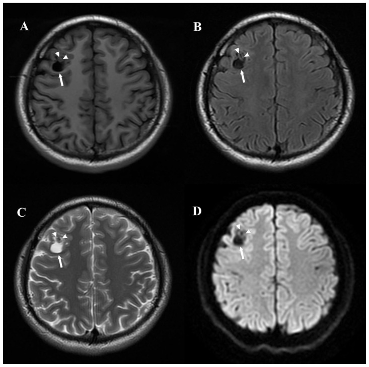
Figure 2.
25-year-old male with angiocentric glioma in the right frontal lobe.
Findings: Axial T1WI (T1-FLAIR) (A), FLAIR (B) and T2WI (C) demonstrate a relatively well defined cystoid lesion (arrow) that is hypo-intense on T1WI and FLAIR image, and hyper-intense on T2WI. There are no diffusion restrictions on DWI (D). The small area of calcification (arrowhead) anterior to the cystoid lesion is isointense on T1WI, hypo-intense on FLAIR image, T2WI and DWI.
Technique: 3.0T Signa HD (GE, USA) MR scanner. 5.0mm slice thickness. T1WI (T1-FLAIR): TR=2580ms, TE=16ms, TI=860ms. FLAIR: TR=9000ms, TE=155ms, TI=2250ms. T2WI: TR=3280ms, TE=108ms. DWI: b-value =0 and 1000 s/mm2, TR=5300ms, TE=75ms.

