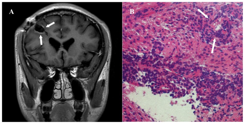
Figure 5.
25-year-old male with angiocentric glioma in the right frontal lobe.
Findings: A. There is no evidence of tumor recurrence on three months follow-up postcontrast TIWI (T1-FLAIR) (arrow). B. Histopathology examination showed that bipolar single cells were centered around the blood vessels (arrow).
Technique: 3.0T Signa HD (GE, USA) MR scanner. 5.0mm slice thickness. T1WI (T1-FLAIR): TR=2580ms, TE=16ms, TI=860ms. Contrast material and dose: Gadolinium 0.2ml/Kg.b) Posterior view shows abnormal increased uptake in the left parotid gland (arrow).

