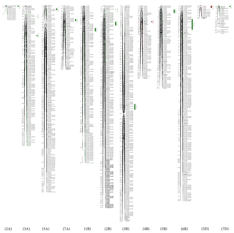
Figure 1.
Segregation distorted region (SDR) detected in the consensus map. The majority of the distorted markers clustered in the SDRs on chromosomes 1B, 2A, 2B, 3A, 3B, 4B, 5A, 5B, 5D, 6B, 7A, and 7D in the consensus map. SSR markers are identified in bold type, and SDRs are labeled with green or brown, indicating SDR bias to the male or female parent, respectively.

