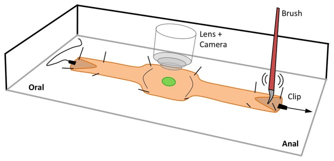
Figure 1.

Schematic of the experimental setup used to record Ca2+ activity in targeted cells in the isolated intact mouse colon. The ends of the preparation were slit and pinned open to allow for mucosal stimulation using a brush and/or longitudinal stretch stimulation using clips. In the middle of the preparation where recordings were taken, the colon was stabilized by pinning through the outer edges.
