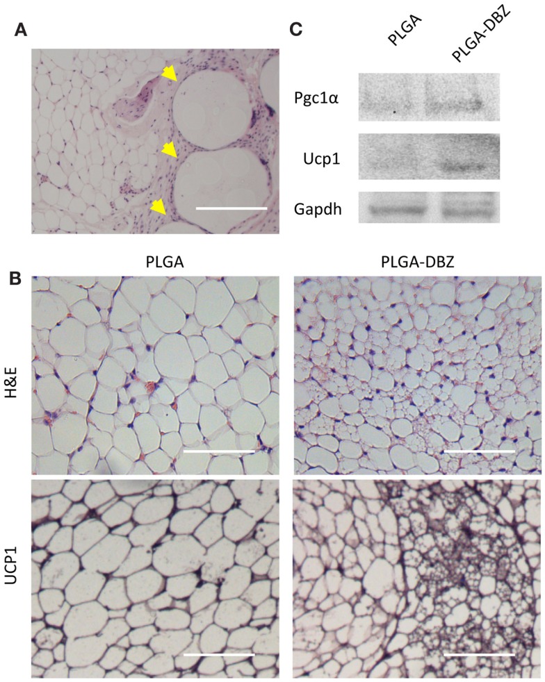
Figure 6.
DBZ-loaded PLGA microspheres induce browning in vivo. (A) H&E staining showing DBZ-loaded PLGA microspheres (yellow arrowhead) dispersed within the WAT depot. Scale bar, 200 μm. (B) H&E (top) and UCP1 (bottom) staining of inguinal WAT from mice at 14 days post-injection of DBZ-loaded PLGA microspheres. Scale bars, 100 μm. (C) Protein levels of Pgc1-α and Ucp1 of inguinal WAT from mice at 14 days post-injection of DBZ-loaded PLGA microspheres.

