Figure 3.
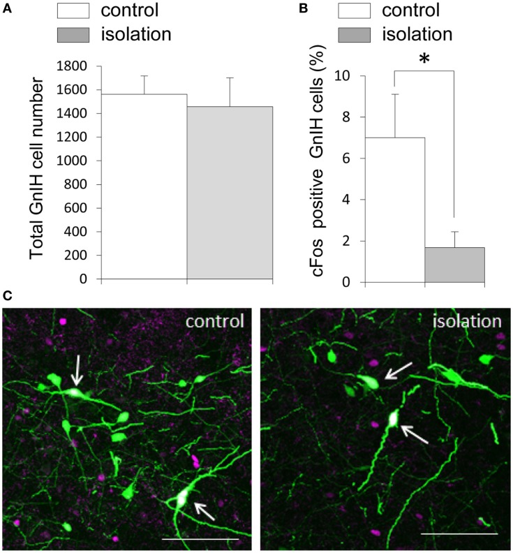
The effect of post-weaning social isolation on enhanced green fluorescent protein–gonadotropin-inhibitory hormone neurons in the dorsomedial hypothalamic nucleus. (A) Total number of gonadotropin-inhibitory hormone (GnIH) cells in the dorsomedial hypothalamic nucleus (DMN) of control and isolated male rats (control = 14 and isolated = 11). (B) Percentage of c-Fos-positive GnIH neurons in the DMN of control and isolated male rats. Data represent the mean ± SEM for each group. *p < 0.05. (C) Confocal images of enhanced green fluorescent protein (EGFP)–GnIH cells expressing c-Fos protein (white, indicated by arrows), EGFP–GnIH neurons (green), and red c-Fos protein (magenta) in control (left panel) and isolated (right panel) male rats. Scale bar: 100 μm.

