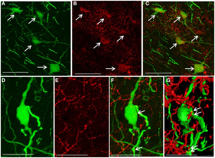
Figure 4.
Co-localization of 5-hydroxytryptamine fibers and the 5-hydroxytryptamine2A receptor in enhanced green fluorescent protein–gonadotropin-inhibitory hormone cells in the dorsomedial hypothalamic nucleus. (A) Enhanced green fluorescent protein (EGFP)–gonadotropin-inhibitory hormone (GnIH) cells and fibers (green), (B) 5-hydroxytryptamine 2A (5-HT2A)-immunostained neurons (red), and (C) EGFP–GnIH cells expressing 5-HT2A (yellow/orange) and 5-HT2A-immunostained neurons (red) in the dorsomedial hypothalamic nucleus (DMN). (D) EGFP–GnIH neurons (green), (E) 5-hydroxytryptamine (5-HT)-immunostained fibers (red), and (F) 5-HT in close juxtapositions in EGFP–GnIH neurons or fibers (yellow dot, indicated by arrows). (G) 3D images of 5-HT in close juxtapositions in EGFP–GnIH neurons or fibers (yellow dot, indicated by arrows) in the DMN. Scale bars: (A–C), 50 μm; (D–G), 20 μm.

