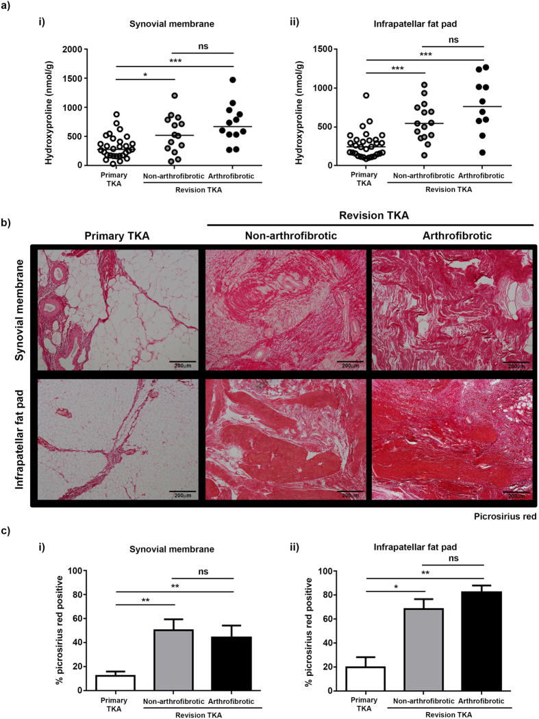
Figure 3. Significant tissue remodelling and collagen deposition following TKA.
(a) In patients where sufficient tissue was available, hydroxyproline was measured as a surrogate for collagen content in tissue. There was a significant increase in the hydroxyproline content of synovial membrane (i) and infrapatellar fat pad (ii) from revision patients compared to primary TKA. There was no significant difference in the hydroxyproline content of either tissue between patients undergoing non-arthrofibrotic revision TKA and patients undergoing arthrofibrotic revision TKA. Synovial membrane; primary TKA n = 29, non-arthrofibrotic revision TKA n = 14, arthrofibrotic revision TKA n = 12. Infrapatellar fat pad; primary TKA n = 28, non-arthrofibrotic revision TKA n = 15, arthrofibrotic revision TKA n = 10. *p < 0.05. ***p < 0.001. (b) Representative images of picrosirius red stained synovial membrane (upper panel) and infrapatellar fat pad (lower panel) from patients undergoing primary TKA (left panel), patients undergoing non-arthrofibrotic revision TKA (middle panel) and patients undergoing arthrofibrotic revision TKA (right panel). There is evidence of significant tissue remodelling characterised by the loss of fat cells and the deposition of large quantities of densely packed collagen fibres in both tissues isolated from patients undergoing revision TKA regardless of clinical diagnosis. Images acquired on a Nikon inverted microscope at 10× magnification. (c) Picrosirius red positive staining was quantified using image analysis software. There was a significant increase in picrosirius red positive staining of synovial membrane (i) and infrapatellar fat pad (ii) from revision patients compared to primary TKA regardless of clinical diagnosis. There was no significant difference in picrosirius red positive staining of either tissue between patients undergoing non-arthrofibrotic revision TKA and patients undergoing arthrofibrotic revision TKA. Synovial membrane and Infrapatellar fat pad; primary TKA n = 9, non-arthrofibrotic revision TKA n = 10, arthrofibrotic revision TKA n = 10. *p < 0.05, **p < 0.01.

