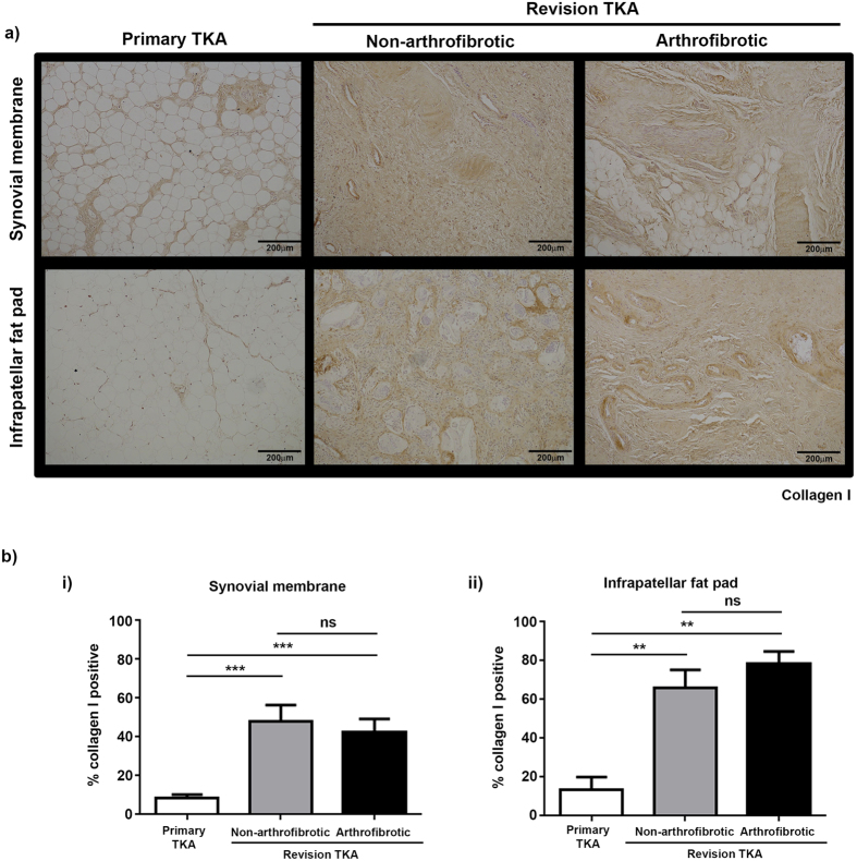
Figure 4. Collagen I is significantly increased in tissue from revision TKA.
Representative images of collagen I stained synovial membrane (upper panel) and infrapatellar fat pad (lower panel) from patients undergoing primary TKA (left panel), patients undergoing non-arthrofibrotic revision TKA (middle panel) and patients undergoing arthrofibrotic revision TKA (right panel). There is a significant increase in collagen I expression in revision TKA tissue compared to primary TKA tissue. Images acquired on a Nikon inverted microscope at 10× magnification. (b) Collagen I positive staining was quantified using image analysis software. There was a significant increase in collagen I positive staining of synovial membrane (i) and infrapatellar fat pad (ii) from revision patients compared to primary TKA regardless of clinical diagnosis. There was no significant difference in collagen I positive staining of either tissue between patients undergoing non-arthrofibrotic revision TKA and patients undergoing arthrofibrotic revision TKA. Synovial membrane and Infrapatellar fat pad; primary TKA n = 9, non-arthrofibrotic revision TKA n = 10, arthrofibrotic revision TKA n = 10. **p < 0.01, ***p < 0.001.

