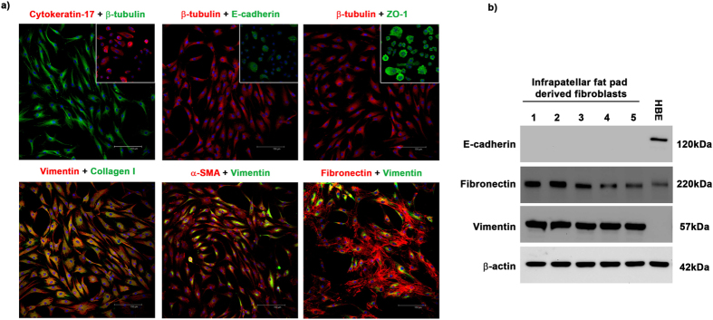
Figure 1. Cells isolated from the infra-patellar fat pad demonstrate a mesenchymal phenotype.
(a) Cells isolated from the infra-patellar fat pad were cultured in vitro and the expression of epithelial and mesenchymal markers investigated by immunocytochemistry. Cells had little to no expression of the epithelial markers cytokeratin 17, E-cadherin and ZO-1. Insets show positive staining on human lung epithelial cells. β-tubulin was used to demonstrate cell morphology. In contrast cells expressed very high levels of the mesenchymal markers vimentin, collagen 1, α-SMA and fibronectin. DAPI was used as a nuclear counter stain. Images were acquired on a Leica TCS SP2 UV confocal microscope at x20 magnification. (b) Whole cell lysates of infra-patellar fat pad derived fibroblasts (n = 5) were investigated for the expression of epithelial and mesenchymal markers by Western blotting. Cells express high levels of fibronectin and vimentin but no E-cadherin. β-actin was used as a loading control. Human bronchial epithelial cells (16HBE14o- (HBE)) were used as a positive control for epithelial marker expression.

