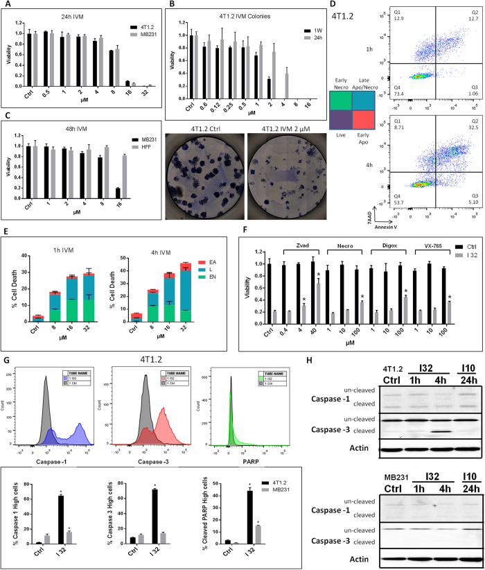
Figure 1. Ivermectin kills breast cancer cells through a mixed apoptotic and necrotic mechanism.
(A) Mouse (4T1.2) and human (MDA-MB-231) TNBC cells manifest similar sensitivity to Ivermectin. Viability of cells treated with various doses of Ivermectin for 24 h. (B) Extended exposure time reduces IC50 values to as low as 2 μM. 4T1.2 cells were seeded at 100 cells/well and individual colonies were counted after a week. Cancer cells were exposed to Ivermectin during the initial 24 h or during the entire duration of the assay. (C) MDA-MB-231 breast cancer cells manifest higher sensitivity to Ivermectin compared to normal non-transformed human foreskin fibroblasts (HFFs). (D) Flow cytometry analysis showing that cell death proceeds through two distinct pathways: a directly necrotic 7AAD-single positive or Annexin V/PS-single positive apoptotic pathway. (E) Kinetics of necrotic versus apoptotic killing of 4T1.2 breast cancer cells. (F) Ivermectin-induced cell death can be reversed by inhibition of various controlled cell death pathways. 4T1.2 cells were treated for 4 h with 32 μM Ivermectin in the presence of μM concentrations of Z-vad-fmk, Necrostatin-1, Digoxin, or VX-765, as indicated. (G) Activation of Caspase-1, Caspase-3 and cleavage of PARP in 4T1.2 and MDA-MB-231 cells treated with 32 μM for 4h. Asterisk (*) indicates p < 0.05 relative to untreated or Ivermectin alone controls, respectively. (H) Western blot analysis showing constitutive and Ivermectin-induced cleavage of caspases 1 and 3 in murine (top) and human (bottom) breast cancer cells.

