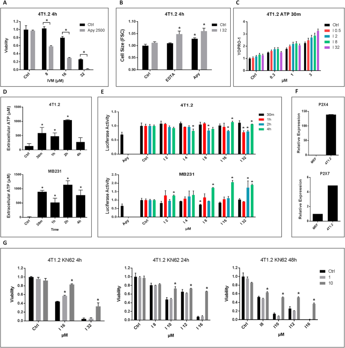
Figure 3. Dual roles of ATP and purinergic signaling in Ivermectin’s killing.
(A) Depletion of extracellular ATP with Apyrase (2500 μunits/ml) exacerbates killing of 4T1.2 cell by Ivermectin. (B) Extracellular Ca2+ and ATP contain Ivermectin-induced cell swelling. 4T1.2 cells were treated with 32 μM Ivermectin for 4 h in the presence of 1 mM EDTA or 2500 μunits/ml Apyrase. (C) Ivermectin (0.5–32 μM) and high concentrations of extracellular ATP (0.3–3 mM) synergistically open pannexin-1 channels and permeabilize the membrane on live cells (7AAD-positive dead cells were gated out). 4T1.2 cells were treated for 30 min as indicated in the presence of 7AAD and 5 μM YOPRO-1. (D) Analysis of supernatants from Ivermectin-treated murine and human TNBC cells showing rapid release of ATP followed by its transient depletion. Depletion of extracellular ATP with Apyrase (2500 μunits/ml) was used as a positive control. (E) Analysis of membrane-proximal ATP levels using cancer cells engineered to express a membrane-bound Luciferase. (F) qPCR demonstrating over-expression of P2X4 and P2X7 receptors in mouse 4T1.2 breast cancer cells versus mouse embryonic fibroblasts (MEF). (G) The P2X7-specific inhibitor KN-62 (1–10 μM) blocks Ivermectin cytotoxicity (IVM 8–32 μM, 4–48 h treatments). Asterisk (*) indicates p<0.05 relative to untreated or Ivermectin alone controls, respectively.

