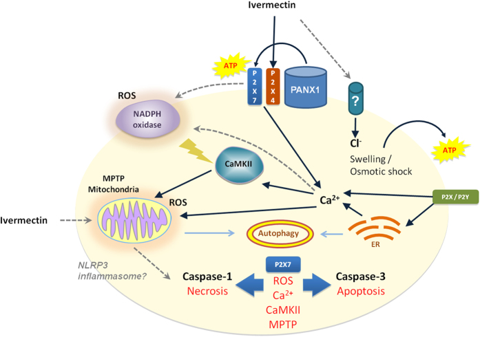
Figure 7. Model of P2X4/P2X7/Pannexin-1-induced cancer cell death.
Ivermectin induces P2X4/P2X7-dependent activation of Pannexin-1 channels and release of ATP. The release of ATP might be transiently protective, but only in cell types that are highly sensitive to Ivermectin-induces cell swelling when ATP and Ca2+ signaling are essential for control of cell volume. In cancer cells where no cell size changes can be observed (for example human TNBC MDA-MB-231 cells), high concentrations of ATP (1–3 mM) immediately enhance Ivermectin cytotoxicity. Potentiated P2X7 receptor signaling drives a fast progressing necrotic/pyroptotic mechanism driven by NADPH oxidases-generated ROS, cytosolic Ca2+/CaMKII activation, and MPTP, and characterized by caspase-1 cleavage, due to possible NLRP3 inflammasome activation. Necrotic killing is followed by a slower progressing apoptotic cell death program mediated by caspase-3 activation. The failure of the default apoptotic pathway might be attributed to faster activation of caspase-1, inadequate autophagic control of mitochondrial MPTP, collapse of cellular energy metabolism, resulting in rapid progression of necrotic cell death. Damage to mitochondria and ER stress as well as potential depletion of cellular ATP reserves simultaneously promote autophagy that might render even the slower apoptotic pathway immunogenic.

